
Digital diagnosis
By Lydialyle Gibson
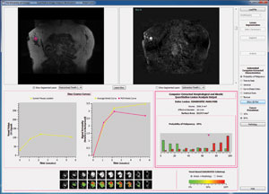
Image courtesy Maryellen Giger
Radiologists use computers to help them identify suspicious regions in mammograms and other cancer-screening images. Called computer-aided diagnosis, the technology acts as a second pair of eyes, leaving the subsequent evaluation—diagnosis, prognosis, and treatment plans—up to the radiologist and oncologist.
Beginning in the mid-1980s, medical physicist Maryellen Giger, PhD’85, and Chicago colleagues Kunio Doi and Heang-Ping Chan, PhD’81, helped develop and commercialize computer-aided detection. “The three of us actually started the field,” says Giger, a professor of radiology and faculty director of the Biological Sciences Division’s Imaging Research Institute.
Now she and her team are creating technology that not only helps radiologists locate potentially cancerous lesions but also uses algorithms to estimate a diagnosis, assess cancer risk, and perhaps predict patients’ response to therapy. Called quantitative image analysis, the system also could be used, she says, to help evaluate patients’ ongoing condition. After beginning treatment, a cancer patient might come in for new imaging every six weeks, she says. Quantitative image analysis would be able to examine the results and help identify, for instance, whether the tumor has shrunk, or if it exhibits other changes in characteristics.
Recently Giger’s team developed a prototype platform technology, which analyzes various types of breast images—mammogram, ultrasound, magnetic resonance—to determine a lesion’s size, the probability of malignancy, and other characteristics. The prototype was selected to be part of the Quantitative Imaging Room at the Radiological Society of North America’s annual meeting in Chicago this past November, where Giger and her lab team gave demonstrations of the workstation (above) and a presentation.Return to top